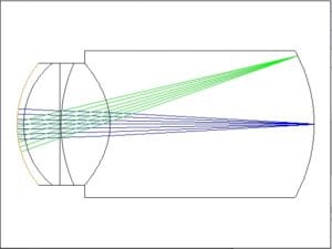
The human eye is a complex optical system that is a bit different for every person and even the same person as they age. This creates challenges for optical engineers trying to simulate and analyze the eye’s performance under different conditions. This blog post delves into the findings from recent analyses of different optical models of the human eye, including the Liou and Brennan model, the Lotmar model, the Navarro model, and others. We will explore key parameters such as image quality, field curvature, distortion, and chromatic aberrations, providing a comprehensive overview of how these models contribute to our understanding of ocular optics.
The Importance of Optical Models
Optical models of the human eye serve as essential tools for understanding how light interacts with the eye’s various components, including the cornea, lens, and retina. These models help researchers and clinicians evaluate visual performance, diagnose refractive errors, and design optical devices such as contact lenses and intraocular lenses. By simulating the eye’s optical properties, we can gain insights into how different factors affect image quality and visual perception.
Overview of Key Optical Models
1. Liou and Brennan Model
The Liou and Brennan human eye model, developed in 1997, is a widely recognized schematic eye model that represents the anatomical and optical characteristics of the human eye with a high degree of accuracy. It includes several essential components of the eye’s structure, such as the cornea, lens, and retina, and captures how these components affect light refraction and focus. Unlike earlier models, Liou and Brennan’s version integrates aspheric surfaces and gradient-index optics within the lens, which more accurately reflects the real eye’s changing refractive index.
This model simulates the way light interacts with the eye, offering enhanced precision in representing visual functions, including the formation of retinal images and the eye’s overall optical aberrations. The Liou and Brennan model is widely used in vision science, optical engineering, and biomedical research, particularly for designing corrective lenses and testing imaging devices.
Key Findings:
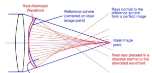
- Image Quality: The model demonstrates diffraction-limited image quality on-axis, with an optical path difference (OPD) of about 0.6 Wave PV for off-axis angles of ±10 degrees and 1.8 Wave PV for ±20 degrees. The Strehl ratio, a measure of image quality, is 0.528 at ±10 degrees and drops to 0.047 at ±20 degrees.
- Field Curvature and Distortion: The model shows a field curvature of approximately 0.02 mm and distortion of about 8%. These parameters are crucial for understanding how the eye focuses light and how image quality degrades off-axis.
- Chromatic Aberrations: The axial color aberration is about 0.08 mm, while the lateral color aberration is approximately 0.002 mm. These aberrations can affect color perception and overall image clarity.
2. Lotmar Model
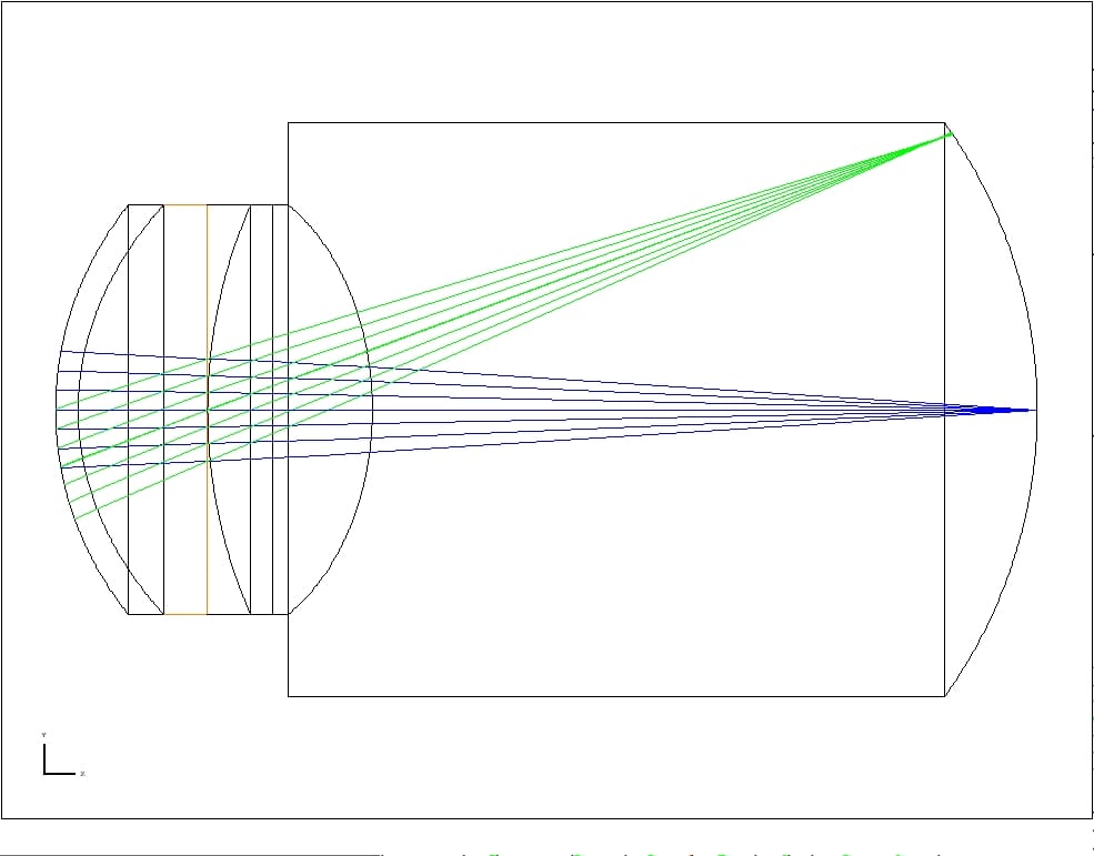
The Lotmar eye model, proposed by Walter Lotmar in 1971, is a simplified representation of the human eye that is particularly useful in studies of retinal imaging and visual optics. It was one of the early attempts to create a mathematically defined eye model that could simulate the optical pathway from the cornea through to the retina. The model divides the eye’s optical system into two primary components: a single refractive surface for the cornea and a crystalline lens with fixed refractive power.
The simplicity of the Lotmar model, as opposed to more complex models like the Liou and Brennan model, makes it accessible for analytical calculations and computational modeling. While the model does not account for aspheric surfaces or gradient-index changes within the lens, it offers a good approximation of how light is refracted and focused onto the retina. The Lotmar model assumes spherical symmetry and incorporates a uniform refractive index within the eye, making it less accurate than later models but valuable for basic studies in retinal imaging, optical aberrations, and vision correction.
Due to its straightforward design, the Lotmar eye model remains useful in educational contexts and in studies where a detailed anatomical model is not essential. However, for more precise applications such as lens design or surgical planning, more advanced models, including the Liou and Brennan model, are generally preferred.
Key Findings:
- Image Quality: The Lotmar model also exhibits diffraction-limited image quality on-axis. However, off-axis performance shows a notable decline, with an OPD of about 1.8 Wave PV at ±10 degrees and greater than 7.0 Wave PV at ±20 degrees. The Strehl ratio reflects this degradation, measuring 0.274 at ±10 degrees and dropping to 0.034 at ±20 degrees.
- Field Curvature and Distortion: The model indicates a field curvature of about 2.5 mm and distortion of approximately 8%. These values highlight the challenges in maintaining image quality across the visual field.
- Chromatic Aberrations: The Lotmar model reports axial color aberration of about 0.22 mm and lateral color aberration of about 0.035 mm, which can impact color discrimination.
3. Navarro Model
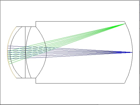
The Navarro eye model, introduced by Rafael Navarro in 1985, is a sophisticated mathematical model of the human eye designed to closely replicate the eye’s actual optical behavior and anatomical structure. It improves upon earlier models by providing a more accurate representation of the cornea, crystalline lens, and other ocular components. Navarro’s model uses aspheric surfaces for both the cornea and lens, which helps simulate real-life aberrations of the eye, such as spherical and chromatic aberrations. Additionally, the model incorporates gradient-index optics within the lens, which more accurately mimics the gradual change in refractive index from the center to the edge of the lens, a feature not present in simpler models.
The Navarro eye model consists of three main components:
- Cornea: Represented by two aspheric surfaces that better approximate the natural shape of the cornea, enabling accurate modeling of light refraction as it enters the eye.
- Crystalline Lens: Uniform refractive index with four refractive surfaces.
- Retina: The model does not directly simulate the retina but instead calculates the quality of the image formed at the retinal plane, simulating how well light focuses onto this plane.
The Navarro model provides a more precise depiction of how light behaves as it travels through the eye, making it suitable for applications in optical research, vision science, and clinical settings. It is particularly valuable in the study of visual optics, designing optical devices such as contact lenses and intraocular lenses, and testing corrective procedures like LASIK. The model’s ability to simulate optical aberrations also makes it useful in evaluating visual performance and understanding the impact of refractive errors.
Key Findings:
- Image Quality: The Navarro model shows diffraction-limited image quality on-axis, with an OPD of about 1.5 Wave PV at ±10 degrees and 6.0 Wave PV at ±20 degrees. The Strehl ratio is 0.149 at ±10 degrees and drops to 0.043 at ±20 degrees, indicating significant off-axis degradation.
- Field Curvature and Distortion: The model indicates a field curvature of about 2.0 mm and distortion of approximately 8%. These parameters are critical for understanding how the eye’s optical system handles peripheral vision.
- Chromatic Aberrations: The axial color aberration is about 0.24 mm, while the lateral color aberration is approximately 0.08 mm, which can affect visual clarity and color perception.
4. Arizona University Model
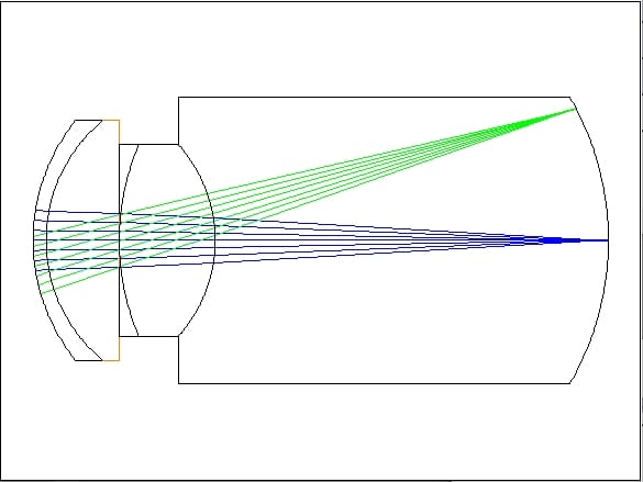
The Arizona Eye Model, developed by researchers at the University of Arizona, is an advanced optical model of the human eye that provides a highly accurate representation of ocular anatomy and optical behavior. This model, which originated in the early 2000s, was designed to support sophisticated research in vision science and optical engineering. It builds upon earlier models, integrating more precise details of the eye’s structural and optical characteristics.
Key features of the Arizona Eye Model include:
- Detailed Anatomical Accuracy: The Arizona model incorporates anatomically accurate data for key eye structures, including the cornea, crystalline lens, anterior chamber, and retina. These components are modeled with aspheric surfaces to closely match the natural curvature and refractive behavior of the human eye.
- Accommodation corrected lens: The Arizona Eye Model includes a single element with witha refractive index that is dependent on the accomodation
- Accommodation Simulation: The model can replicate the eye’s accommodation process, or the ability to change focus between near and far objects. By adjusting the curvature of the lens surfaces and the gradient index, the model can simulate changes in the eye’s focusing power, making it useful for studies of presbyopia and other accommodation-related vision issues.
- Modeling of Aberrations: The Arizona Eye Model is designed to accurately predict optical aberrations, including spherical, chromatic, and higher-order aberrations. These aberrations are critical in understanding visual clarity and designing corrective optics, such as contact lenses and intraocular lenses.
- Versatility for Research and Clinical Applications: The model is used in a range of applications, from developing corrective lenses to enhancing surgical procedures and simulating the impact of various ocular conditions on vision. Its high level of anatomical and optical detail also makes it a valuable tool in evaluating the performance of optical devices under realistic conditions.
The Arizona Eye Model is widely respected for its detailed and realistic representation of the eye, providing a foundational tool in fields like ophthalmology, visual optics, and biomedical engineering. Its flexibility to simulate various optical conditions, combined with its accuracy in representing human eye optics, makes it a preferred model for both academic research and practical applications.
Key Findings:
- Image Quality: The Arizona model demonstrates excellent on-axis performance, with an OPD of less than 0.1 Wave PV and a Strehl ratio of 0.987. Off-axis performance shows an OPD of about 0.5 Wave PV at ±10 degrees and greater than 2 Wave PV at ±20 degrees, with a Strehl ratio of 0.162.
- Field Curvature and Distortion: The model indicates a field curvature of about 0.02 mm and distortion of approximately 3%. These values suggest that the Arizona model maintains better image quality compared to other models.
- Chromatic Aberrations: The axial color aberration is about 0.25 mm, while the lateral color aberration is approximately 0.035 mm.
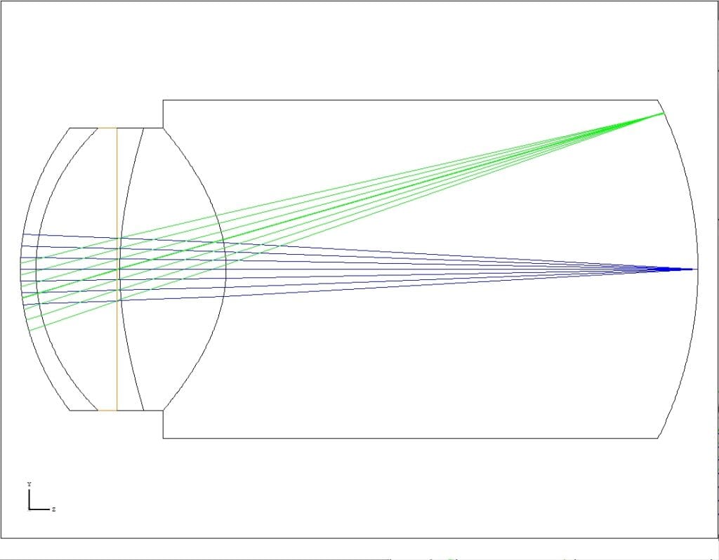
Comparative Analysis of Optical Models
When comparing the various optical models, several key observations emerge:
Image Quality
All models exhibit diffraction-limited image quality on-axis, but off-axis performance varies significantly. The Liou and Brennan model shows the least degradation at ±10 degrees, while the Navarro and Lotmar models experience substantial declines in image quality at larger angles. The Arizona model stands out for its superior on-axis performance and relatively better off-axis quality.
Field Curvature and Distortion
Field curvature and distortion are critical parameters that affect image quality. The Lotmar and Navarro models report higher field curvature values, which can lead to more significant image distortion. In contrast, the Arizona model demonstrates lower distortion, suggesting a more favorable optical design.
Chromatic Aberrations
Chromatic aberrations are essential for understanding color perception. The Liou and Brennan model exhibits the lowest axial color aberration, while the Navarro model shows the highest. This variation can impact how different models are applied in clinical settings, particularly in designing optical devices.
Here’s a comparison of the Liou and Brennan, Lotmar, Navarro, and Arizona eye models, summarizing their key features, complexity, and common applications. This table highlights the evolution from simpler to more anatomically and optically accurate models, with each model bringing unique strengths for specific research and clinical uses
| Feature | Lotmar Eye Model | Liou and Brennan Eye Model | Navarro Eye Model | Arizona Eye Model |
| Year Developed | 1971 | 1997 | 1985 | Early 2000s |
| Model Structure | Simplified, single refractive lens | Anatomically accurate, aspheric surfaces | Anatomically accurate, aspheric surfaces | Highly detailed, anatomically accurate |
| Corneal Representation | Spherical surface | Aspheric, realistic curvature | Aspheric, realistic curvature | Aspheric, with high anatomical accuracy |
| Crystalline Lens | Single index, no gradient | Gradient-index | Single index | Multiple indexes |
| Gradient-Index (GRIN) Lens | No | Yes | No | No |
| Aberrations Modeled | Basic | Spherical, chromatic | Spherical, chromatic, higher-order | Spherical, chromatic, higher-order |
| Accommodation Simulation | No | Limited | Yes | Yes, detailed |
| Applications | Educational, basic studies | Visual optics, corrective lens design | Advanced optical research, device testing | Clinical research, surgical planning |
| Strengths | Simplicity, ease of calculations | Anatomical realism, GRIN lens | High optical realism, realistic aberrations | Highest accuracy, full accommodation simulation |
| Limitations | Limited realism, lacks aspheric surfaces | Moderate accommodation simulation | Limited anatomical detail relative to Arizona model | High complexity, computationally intensive |
Implications for Optical Design
Which optical model the system designer selects can have significant implications for the design of optical devices, including contact lenses, intraocular lenses, and retinal cameras. Understanding how different models perform under various conditions allows researchers and clinicians to understand how different optical devices might perform.
Designing Optical Devices
- Contact Lenses: Knowledge of how the eye’s optical system behaves can guide the design of contact lenses that minimize aberrations and improve overall visual quality. By selecting materials and geometries that align with the eye’s optical properties, manufacturers can create lenses that enhance visual performance.
- Intraocular Lenses: For patients undergoing cataract surgery, the choice of intraocular lens (IOL) is critical. The insights from optical models can help surgeons select IOLs that provide optimal image quality and minimize aberrations, leading to better postoperative outcomes.
- Retinal Cameras: The design of retinal cameras relies heavily on understanding the eye’s optical properties. By simulating the eye’s performance using these models, engineers can create cameras that capture high-quality images of the retina, aiding in the diagnosis and monitoring of ocular diseases.
Conclusion
The exploration of various optical models of the human eye provides valuable insights into the complex interactions between light and the eye’s components. By analyzing parameters such as image quality, field curvature, distortion, and chromatic aberrations, researchers can enhance our understanding of ocular optics and improve the design of optical devices. As technology continues to advance, these models will play an increasingly vital role in the field of optometry and ophthalmology, ultimately leading to better visual outcomes for patients.
For more information on optical modeling and the latest research in ocular optics, visit [www.opticsforhire.com]
FAQ on Eye Models
1. What is the primary purpose of these eye models?
These eye models simulate the optical and anatomical properties of the human eye to study how light interacts with its components. They are used in vision science, optics, and clinical research for applications such as lens design, retinal imaging, and studying visual aberrations.
2. How do the models differ in complexity?
- Lotmar Eye Model: Simplified and easy to use; suited for basic studies and educational purposes.
- Liou and Brennan Model: More realistic, featuring aspheric surfaces and a gradient-index lens, allowing for moderate simulation of optical properties.
- Navarro Eye Model: High accuracy with gradient-index optics and aspheric surfaces, modeling aberrations effectively.
- Arizona Eye Model: The most detailed and realistic, capable of simulating accommodation and highly accurate optical behavior.
3. Which model is the best for studying optical aberrations?
The Navarro Eye Model and Arizona Eye Model are best for studying optical aberrations due to their aspheric surfaces and gradient-index lens representation. The Arizona model offers the most comprehensive analysis, including higher-order aberrations.
4. Can any of these models simulate accommodation?
Yes, the Navarro Eye Model and Arizona Eye Model can simulate accommodation:
- Navarro: Offers moderate accuracy in simulating focus changes.
- Arizona: Provides highly detailed accommodation simulation, making it ideal for presbyopia studies and lens design.
5. Why is the Lotmar Eye Model still used despite its simplicity?
The Lotmar Eye Model is simple, computationally inexpensive, and suitable for basic studies or teaching purposes. It is a good starting point for researchers or students learning about retinal imaging and basic refractive properties of the eye.
6. How does the Liou and Brennan Model improve upon the Lotmar Model?
The Liou and Brennan Model adds anatomical realism with aspheric surfaces for the cornea and gradient-index optics in the lens. These features allow it to simulate more realistic light refraction and basic aberrations.
7. What makes the Arizona Eye Model unique?
The Arizona Eye Model is the most anatomically accurate and versatile, capable of simulating accommodation, optical aberrations, and the effects of surgical interventions. It is widely used in advanced research and clinical applications, such as designing corrective lenses and planning eye surgeries.
8. Which model is suitable for designing corrective lenses?
The Liou and Brennan, Navarro, and Arizona models are all suitable for designing corrective lenses:
- Liou and Brennan: Effective for basic optical corrections.
- Navarro: More advanced lens testing, including aberration correction.
- Arizona: The best for detailed and precise corrective designs.
9. Are these models computationally demanding?
- Lotmar: Not computationally demanding; simple calculations.
- Liou and Brennan: Moderately demanding.
- Navarro: Requires more computational resources for advanced aberration analysis.
- Arizona: Highly computationally intensive due to its anatomical detail and accommodation simulation.
10. Can these models simulate individual differences in eyes?
Most models are generalized representations of the human eye, but the Arizona Eye Model provides the flexibility to adjust parameters for individual differences, making it suitable for personalized studies and clinical research.

